For improved readability, to make text smaller or larger, click the buttons below
How to Reach Us
Our address, phone number, and location
Bozdech Eye Clinic
#120, 5002-55 Street
Red Deer, Alberta
T4P 7A4
P: 403-342-2020
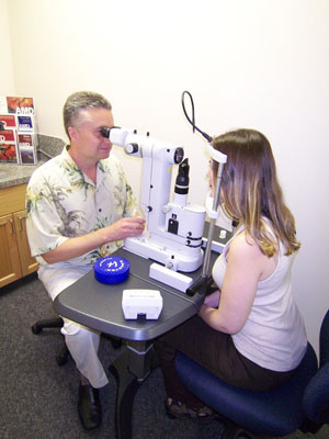
Eye Pre-Exam Instructions
Important things to know before your eye exam
Please bring:
- A list of medications you are currently taking.
- If you are a diabetic, bring along a snack or any medications you may need, in case of increased waiting time.
- Sunglasses, for your comfort post-procedure.
- Your corrective eye wear, e.g. glasses, contact lenses.
- Someone to drive you, or plan an alternate mode of transportation to return home.
What to expect:
- Your appointment is expected to last approximately 1 to 2 hours, including dilation, vision screening and consultation.
- Dilation can last from 4 to 6 hours after your appointment, and while your eye(s) are dilated, we do not recommend operating a motor vehicle. Therefore, we suggest that you bring someone to drive you, or plan an alternate mode of transportation to return home.
- We suggest that you do not bring small children with you to your appointment unless absolutely necessary.
Eye Conditions & Treatments
Learn more about eye conditions we treat
Diabetes Read More
Did you know that diabetes is the leading cause of blindness in Canada in people under 60 years of age?
Over 3.4 million Canadians have this disease, and about half of them experience damage to their vision. Diabetes can lead to eye conditions such as cataracts, double vision, glaucoma and even blindness. An important cause of visual loss in diabetics is diabetic retinopathy and swelling of the retina called macular edema. The longer you are a diabetic, or the poorer your blood sugar control, the greater your risk of developing diabetic retinopathy. Diabetics also run the risk of developing other complications such as kidney failure, heart attacks and strokes. Research has shown that controlling blood glucose levels, blood pressure and blood cholesterol or lipid levels slows down or even halts diabetic complications.
Diabetic retinopathy involves a change in the blood vessels that feed the retina lining the back of the eye. These blood vessels weaken and leak fluid or blood. Sometimes the retina sprouts new abnormal vessels. This causes swelling of the retina and can lead to retinal or optic nerve infarction (stroke). The eye can fill up with blood or the retina can detach altogether.
It is therefore very important to be monitored by an ophthalmologist on a regular basis, at minimum annually. At our clinic we have the expertise and equipment to deal with any diabetic eye complications before they lead to visual loss. Ideally, timely intervention can be provided before vision loss occurs, at which point it may be too late for any chance of visual recovery. Such intervention may consist of change in medications, identification and treatment of concurrent aggravating conditions and risk factors, as well as retinal laser treatment, intraocular drug injections and surgery.
Dr. Bozdech has a special interest in diabetic eye disease. At Bozdech Eye Clinic we hold specific scheduled diabetic clinic days in order to improve office efficiency and provide better service.
Cataract Surgery Read More
A cataract is a clouding of the lens of the eye, through which light enters the eye. When normal, the lens is clear and is located just behind the iris (coloured part of the eye). There are many causes for cataracts.
These include:
- age
- genetics
- trauma
- diabetes
- sun exposure
- cigarette smoking
- certain drugs such as steroids
Cataracts are treated with surgery as neither medications nor glasses can correct this medical problem.
Can cataracts come back? No, cataracts cannot regrow. However, sometimes the posterior capsule becomes opaque and needs to be divided with the YAG laser. This usually only occurs once, so you will not need to have this done again.
When Dr. Bozdech performs cataract surgery he generally uses Alcon's AcrySof SN60WF artificial foldable lens, which had been shown to reduce the risk of postoperative posterior capsular opacification as compared to other implants available on the market. Furthermore, the AcrySof SN60WF lens is specially designed to reduce the harmful effects of visible and ultraviolet light on the retina. This is particularly important in the prevention and progression of macular degeneration. Some patients receive a Toric lens to correct astigmatism; however this lens procedure is not covered by health care and has additional costs.
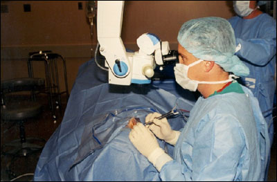
Dr. Bozdech routinely utilizes the most modern technique in cataract surgery, clear corneal surgery under topical anesthesia. This means that most cases are performed under topical anesthesia, consisting of numbing drops and ointment only. In other words, it is a "no needle surgery", unless circumstances dictate otherwise. This completely prevents needle associated complications such bleeding behind the eye and needle associated discomfort, inadvertent ocular perforation leading to blindness, reduced vision or double vision by damaging the muscles/nerves controlling the eye movements.
During cataract surgery Dr. Bozdech makes two very small corneal incisions: one is 2.75 mm and the other is approximately 1 mm. Both incisions are made through the cornea to avoid blood vessels and prevent conjuctival scarring. Sometimes this approach can reduce prexisting corneal astigmatism. Another advantage is that this surgery can be performed successfully in people with bleeding disorders or on blood thinners in the event that they cannot discontinue their medications prior to surgery.
Below are a few videos describing cataracts and what to expect in surgery.
Glaucoma Read More
Glaucoma is an eye disease usually caused by increased pressure within the eye. This disease affects 1 in 100 Canadians over the age of 40. By having regular eye exams the onset of this disease can be prevented.
There are different types of glaucoma, the most common of which is open-angle glaucoma.
Open-angle glaucoma: symptoms can go unnoticed until it is very advanced at which point it may be too late. The reason for this is that it takes out a person's side vision slowly. In adults, this vision loss is permanent. Early intervention is important to save what vision is left. Furthermore, the more advanced the disease becomes the more difficult it is to control.
Treatment options for this disease include eyedrops, which is the most common treatment in North America. Other treatments include surgery and laser treatment. To learn about the laser treatment of glaucoma please click the following link: "SLT Laser". Dr. Bozdech has this special laser on site.
Closed-angle glaucoma is less common: only an eye care professional can tell if you have the eye structure that may lead to this problem. Symptoms of acute closed-angle glaucoma include sudden dull, aching pain over one eye, blurring of the vision or seeing haloes around lights. If you have sudden onset of these symptoms, please seek immediate attention from your doctor. It is treated with a combination of medical and laser therapy.
For more information please click on the following link. GetEyeSmart.org
See our Glaucoma Brochure (PDF):
Macular Degeneration Read More
Age-Related Macular Degeneration (ARMD) is a chronic degenerative disease of the macula. The macula allows you to see fine details directly in front of you as opposed to side vision. There are two types of ARMD, a dry and a wet type.
Dry ARMD progresses slowly and is less severe than the wet type. The wet type consists of abnormal leaky blood vessels. Both types can damage the retina and can take away your central vision. There are many risk factors.
Risk factors that you cannot control include:- Age
- Family History
- Gender: women are more susceptible.
- Smoking
- Diet
- Excessive exposure to sunlight
- High Blood Pressure
- Obesity
Treatment of dry ARMD is focused on monitoring and slowing progression of the disease. This is mainly accomplished by risk factor modification such as quitting smoking, wearing sunglasses on sunny days and supplementing your diet with vitamins such as Vitalux and omega-3 fatty acids.
Omega-3 fatty acid supplementation comes in two forms: fish oils or long-chain fatty acids, and flax seed oil or short-chain fatty acids. Both forms differ somewhat in their effects. The simplest thing is to take both at least twice weekly, not exceeding 3 grams per day to be safe. Since most people with ARMD are already taking Vitalux or an equivalent which contains high doses of vitamin A, one must be careful not to take fish oil capsules derived from fish liver oil as well. These also contain vitamin A, which may lead to vitamin A overdose. Thus take fish capsules derived from fish body oil instead. Patients on blood thinners must be aware that fish oils may enhance the blood thinner effect. However, the flax seed version is safe to be taken with blood thinners. Furthermore, omega-3 oils can disrupt LDL cholesterol and blood sugar control in diabetics. If in doubt about your supplements, ask your pharmacist for help or bring your bottles to Dr. Bozdech.
Wet ARMD is generally treated with anti-VEGF (Vascular Endothelial Growth Factor) therapy such as repeat injections of a substance called Avastin, Eyelea or Lucentis into the eye. Avastin is not covered by health care but Lucentis is covered by some Blue Cross plans. Eyelea (Aflibercept) is a new drug recently approved for use in Canada. Dr. Bozdech can provide these treatments in the office.
When vision becomes significantly impaired, Dr. Bozdech will refer you to the Canadian National Institute of the Blind (CNIB), where expert counselling is provided and various visual aids such as magnifiers can be custom fitted.
For more information visit: amdsupport.ca or GetEyeSmart.org
Retinal Detachment Read More
If immediate treatment is not received, retinal detachment can cause severe permanent loss in your vision.
In some cases there may be small areas of the retina that are torn. These areas, called retinal tears or retinal breaks, which in turn can lead to retinal detachment. When the retina detaches, it is lifted or pulled from its normal position.
Retinal detachment can occur at any age, but it is more common in people over age 40. It affects men more than women, and light-skinned more than dark-skinned people.
A retinal detachment is also more likely to occur in people who:- are extremely nearsighted
- have had an ocular injury
- have a family history of retinal detachment
- have had cataract surgery
- have other eye diseases or disorders, such as retinoschisis, uveitis, degenerative myopia, or lattice degeneration
- have had a previous retinal detachment in the other eye
Retinal detachment can be treated with laser surgery. Retinal detachments may also require surgery to return the retina to its proper position in the back of the eye. A gas bubble is often injected into the vitreous space and is used to push the retinal tear back, thus closing it. Vitrectomy may be necessary to remove any vitreous gel which is pulling on the retina. A scleral buckle may also be placed in the eye, to help hold the retina in place.
Floaters & Flashing Lights Read More
Small specks or clouds moving in your field of vision are called floaters.
When people reach middle age, the vitreous gel may start to thicken or shrink, forming clumps or strands inside the eye. Floaters often occur when the vitreous gel pulls away from the back wall of the eye, causing a posterior vitreous detachment. In some cases the retina can tear if the shrinking vitreous gel pulls away from the wall of the eye. A torn retina is always a serious problem, since it can lead to retinal detachment.
When the vitreous gel inside your eye rubs or pulls on the retina, you may see what look like flashing lights or lightning streaks. The flashes of light can appear off and on for several weeks or months. As we grow older, it is more common to experience flashes. While not all floaters and flashes are serious, you should always have a medical eye examination by an ophthalmologist to make sure there has been no damage to your retina.
Dry Eye Read More
Dry eye syndrome is a very common condition. Although aging certainly plays a key role in reducing natural tear production (especially during menopause), dry eye can affect anyone.
Tear film is made up of a mucous layer against the eye, a middle aqueous (water) layer, and an outer lipid (oily) layer. All three components are important to a normal tear film. If any of the three layers of the tear film are deficient or dysfunctional, the eye may suffer symptoms of dry eye.
Complaints of dry eye may include burning, stinging, redness of the eyes and tearing. The reason why tearing can also be a symptom of ocular dryness is because tear film can be unstable like a drop of water in a greasy frying pan and run off the surface instead of sticking to the eye and keeping it wet. In other words ocular dryness is a complex problem and is often multifactorial.
Various conditions causing dry eyes include aging, hormonal changes (menopause), smoking, medications (blood pressure, antihistamines, antidepressants, birth control pills), contact lens wear, previous refractive laser eye surgery, lack of blinking due to conditions like Parkinson's Disease, or prolonged staring at a computer screen.
Treatment depends on the cause and mechanism of the dry eye syndrome which should be first properly diagnosed by an ophthalmologist. Why see a medical specialist for this? That is because many ocular conditions even as seemingly benign as dry eye can be as a result of an undiagnosed or poorly treated underlying medical condition, such as collagen-vascular disease, or other coexistent ocular conditions such as allergies, rosacea or blepharitis. Furthermore, dry eyes can lead to complications leading to visual loss.
Treatment modalities include removal of the cause e.g. changing medication and quitting smoking. Sometimes dietary changes such as increasing omega-3 fatty acid intake (fish and flax seeds), artificial tear replacement drops and punctal occlusion are also helpful. One can also consider obtaining a humidifier, wearing sunglasses in windy conditions and trying to blink more often. More severe cases can be treated with punctum plugs or Cyclosporin A drops.
Strabismus Read More
Surgery is required when there is ocular misalignment such as crossing over of the eyes (esotropia) or outward pointed eyes (exotropia).
When one eye is pointing up with respect to the other then that is called hypertropia. One may experience double vision when this occurs. In infants this is usually due to congenital abnormalities in the visual pathway or brain development and people sometimes refer to it as a lazy eye. In adults there are many possible causes including stroke, inflammation, trauma and cancer.
To fix strabismus, surgery is sometimes used to realign the muscles that control the eye alignment and movement. The need for surgery depends on which way the eyes are turning, the severity of the turned or crossed eyes, and whether or not improvements can be made through glasses or vision therapy.
Dr. Bozdech performs these surgeries under general anesthesia and the surgery takes about one hour. More complicated cases are generally referred to strabismus specialists in Calgary or Edmonton.
Blepharoplasty Read More
Blepharoplasty is a procedure in which fat is removed, along with excess skin and muscle, from the upper and lower eyelids.
Other Procedures Read More
Removal of eye lid lesions (lumps and bumps), surgical treatment for some tearing disorders, drainage of abscesses and chalazions, eyelid reconstructions, pterygium removal and perforated or lacerated eye repairs to name a few.
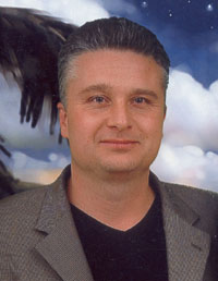
Dr. M. B. Bozdech, B.Sc., M.D., FRCSC
Ophthalmologist & Eye Surgeon
Dr. Bozdech is a fifth generation medical doctor who was born in Czechoslovakia and immigrated to Canada as a child. He received his B.Sc. in Cellular, Molecular and Microbial Biology from the University of Calgary in 1988, his medical degree from the University of Saskatchewan in 1996 and his ophthalmology specialty certification FRCSC (Fellow of The Royal College of Physicians and Surgeons of Canada) in 2001. He received the American Academy of Ophthalmology International Education Award in 2009 and an International Scholar Award in 2010.
Dr. M. Bozdech and his family moved to Grande Prairie in 2001, where he began his career as an ophthalmologist and eye surgeon. In 2004 he spent one year as an appointed Chief of Surgery at the Queen Elizabeth II Hospital. He served as the president of Peace Country Health Regional Medical Organization for 5 years while continuing his practice. Dr. Bozdech relocated to Red Deer in 2012.
Dr. Bozdech also served as a regular member with the Royal Canadian Mounted Police from 1988 to 1992.
Dr. Bozdech’s professional affiliations include:
- Royal College of Physicians and Surgeons of Canada
- College of Physicians and Surgeons of Alberta
- American Academy of Ophthalmology
- Canadian Ophthalmological Society
- Ophthalmology Society of Alberta
- Canadian Medical Association
- Alberta Medical Association
- Royal Canadian Mounted Police Veteran’s Association
- Red Deer Lion's Club
In solo practice, Dr. Bozdech treats conditions such as ocular and periocular traumas, ruptured/perforated eyes, lacerated lids and other complicated eye conditions. He monitors, diagnoses and treats a broad spectrum of eye diseases such as glaucoma, diabetic complications and cataracts. As a medical doctor, he can order MRIs, CT scans and other diagnostic tests anywhere in Canada.
Dr. Bozdech limits his practice to the treatment of medical conditions and therefore does not perform corrective laser eye surgeries, nor does he normally prescribe glasses and contact lenses, except in extenuating circumstances if patients cannot obtain these services to their satisfaction from their local optometrists. The laser treatments Dr. Bozdech performs are only for medical conditions such as posterior capsular opacifications, diabetes, vascular abnormalities, retinal tears and glaucoma.
Dr. Bozdech performs surgeries mostly in Innisfail and Wetaskiwin hospitals and occasionally in the Red Deer hospital.
Our clinic is a proud sponsor of the Red Deer Hospital Foundation and Cross Country Canada athletic development through Project Podium.
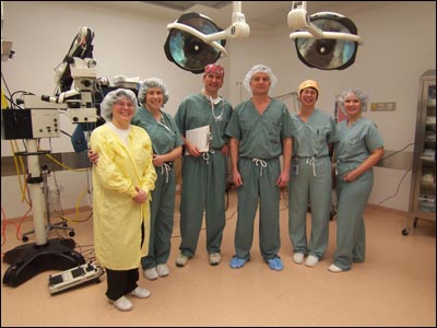
Our Surgical Facilities
Where we perform operations
Dr. Bozdech has surgical privileges at three hospitals: Red Deer, Innisfail and Wetaskiwin.
All three hospitals have excellent nursing staff and equipment and are accessible to patients from out of province. Provincial medicare coverage, from all provinces in Canada, is honoured at all of our facilities.
For cataract surgery, foldable AcrySof lenses may not be covered in some provinces, but they are covered for out of province patients in our surgical facilities via reciprocal agreement. In other words, patients with out of province medical coverage will not have to pay extra for their foldable intraocular lens implants, as they may be required in their home provinces.
Dr. Bozdech also performs some minor eye and eyelid surgeries at his clinic, in the Red Deer hospital outpatient department and at the Innisfail surgical center.
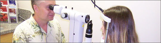
Clinical Equipment
Learn more about the tools and technologies we use
Humphrey Field Analyzer Read More
The purpose of the Humphrey automated field analyzer is to aid in the diagnosis of ocular diseases, most commonly glaucoma. It is also helpful in evaluating and diagnosing neurological diseases such as brain tumors and compressive aneurysms.
The Humphrey offers many different types of testing. When deciding which method of testing is most appropriate, Dr. Bozdech will take several factors into consideration:
- Age
- Family History
- Patient Complaint
- Degree of co-operation
Dr.Bozdech's office is equipped with 2 Humphrey Visual Field Analyzers.
OCT Unit Read More
The Ocular Coherence Tomography (OCT) unit is a state of the art Zeiss non-invasive device capable of scanning the retina and optic nerves in a patient's eye within 10 micron resolution.This device is available at our eye clinic free of charge as it is covered by Alberta and British Columbia health care benefits. The OCT helps Dr. Bozdech objectively analyze and monitor the structures of the eye. It helps with the diagnosis and monitoring of glaucoma, age-related macular degeneration, macular edema, central serous chorioretinopathy, epiretinal membrane and other diseases.
The below PDF brochure gives more information on the OCT unit.
View Flash Video/Presentation - Requires Flash Media Player to view.
SLT Laser Read More
Selective Laser Trabeculoplasty (SLT) is one of the greatest advances in the surgical treatment of glaucoma and is available at the Bozdech Eye Clinic. Compared to the previously utilized technology, argon laser trabeculoplasty (ALT), it is less invasive and just as effective. This repeatable treatment promotes cellular regeneration without the burn and scar tissue associated with other laser procedures.
IOL Master Read More
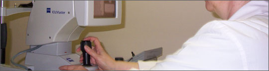
The IOL Master is a scanning device that is used to determine the power of an intraocular lens implant prior to cataract surgery. This state-of-the-art laser system evaluates the length of the eye, surface curvature and anterior chamber depth with great accuracy. With this information it then calculates the intraocular lens power. The information obtained from the IOL Master will allow Dr. Bozdech to replace the eye's natural lens with the greatest precision possible on the market. There is a greater chance of the patient being able to see without glasses for distance vision or reading, depending on patient's choice. If one chooses distance vision, then reading glasses will most likely still be needed. Dr. Bozdech does not recommend multifocal lens as he believes they are associated with a degree of visual compromise and greater potential for patient dissatisfaction.
What other options are there to the IOL Master? Traditionally some ophthalmologists still use ultrasound to accomplish same thing. Ultrasound requires a placement of a probe onto ocular surface (touches the eye) and therefore is invasive and less accurate. It can cause a corneal abrasion. Dr. Bozdech also has such a device but only uses it for special cases where cataracts are so dense that the laser light from IOL Master cannot accurately take the measurement.
Other Lasers Read More
Nd:YAG Laser stands for Neodymium-doped Yttrium-Aluminum-Garnet Laser. Dr. Bozdech uses this in his office to remove a posterior subcapsular opacification (scar) that sometimes develops after cataract surgery. It causes blurry vision that generally clears up with laser.
Argon Laser: Dr. Bozdech uses this laser system to treat retinal tears in order to prevent retinal detachment/visual loss. It is also used to treat proliferative diabetic retinopathy and macular edema (retinal swelling).
These laser treatments are available in our office and are covered by Alberta Health Care.
Ultrasound Read More
Dr. Bozdech has 3 types of ocular ultrasounds in his office:
Axis A-scan is used for measuring the size of eyeballs with dense cataracts for the purpose of calculating the intraocular lens implant power needed for cataract surgery, in order to attain reasonable postoperative spectacle independence.
Accutome B-scan is an instrument that allows Dr. Bozdech to evaluate intraocular and orbital structures when the view of the retina is obscured by blood or other media opacities. It can be useful in diagnosing and monitoring intraocular tumors or other lesions.
UBM (ultrasound biomicroscopy) is a highly specialized ultrasound which allows evaluation of certain intraocular structures and their relationships with respect to each other. For example, this instrument allows more accurate diagnosis of angle closure glaucoma or predisposition for this blinding disease. It is the only way to properly visualize and diagnose any lesions hiding immediately behind the iris.
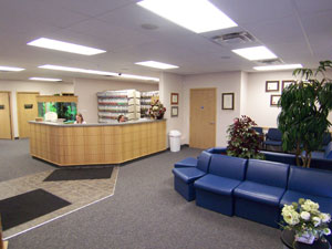
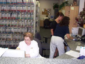
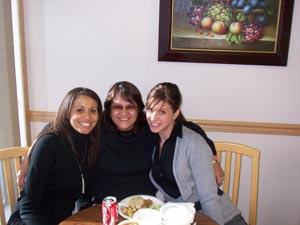
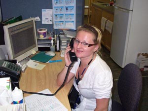
Photo Gallery
Pictures of our clinic and helpful staff